Quick Answer: A post-meal blood sugar of 117 mg/dL is within normal range and aligns with healthy glucose control, especially when considering timing differences with other readings.
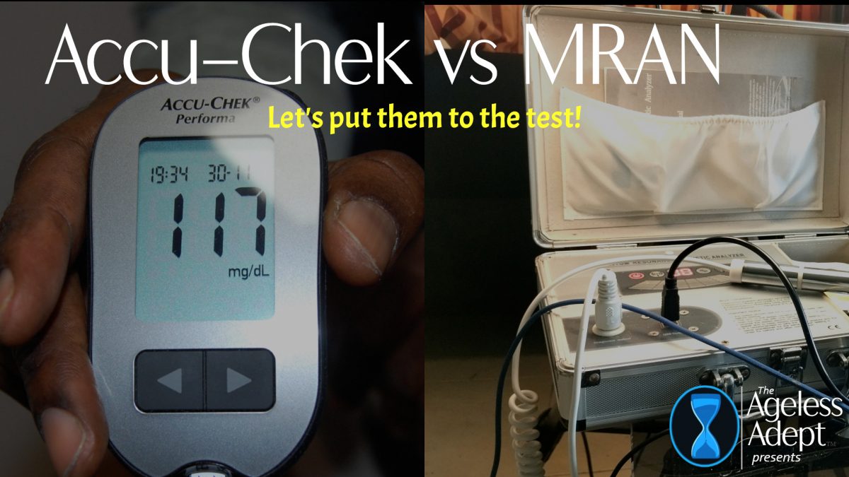
Using an Accuchek Meter at a local eye clinic, my blood sugar level was determined to be 117mg/dL. The test was administered about an hour and a half after eating a cereal-almond milk-raisin-banana-bee pollen breakfast. What can this tell me about my diabetes status?
![]() A blood sugar level of 117 mg/dL about 1.5 hours after eating (postprandial) falls within the normal range for most people, especially for someone without diabetes. Here’s what it might indicate:
A blood sugar level of 117 mg/dL about 1.5 hours after eating (postprandial) falls within the normal range for most people, especially for someone without diabetes. Here’s what it might indicate:
Normal Blood Sugar Ranges (per ADA guidelines):
-
Fasting (before eating): 70–100 mg/dL (normal); 100–125 mg/dL (prediabetes); 126 mg/dL or higher (diabetes).
-
1–2 hours after eating (postprandial): Less than 140 mg/dL (normal); 140–199 mg/dL (prediabetes); 200 mg/dL or higher (diabetes).
What Your Reading Suggests:
-
Your reading of 117 mg/dL suggests that your blood sugar is well-controlled after eating. This value is within the normal postprandial range for non-diabetic individuals.
-
The meal you consumed (cereal, almond milk, raisins, banana, and bee pollen) includes a mix of carbohydrates and natural sugars, which can raise blood sugar levels. However, your body’s ability to handle this indicates no immediate concern for diabetes or impaired glucose metabolism.
What You Can Do:
-
Monitor Trends:
-
A single reading isn’t enough to determine your diabetes status. Regularly monitor fasting and postprandial blood sugar over a few weeks to identify patterns.
-
-
Consider an A1C Test:
-
If you’re curious about your overall blood sugar control over the past 2–3 months, you can request a hemoglobin A1C test from your doctor.
-
-
Meal Composition:
-
If you’re keeping your blood sugar stable, continue focusing on balanced meals with fiber, healthy fats, and protein to moderate blood sugar spikes.
-
Assuming the blood drop-based test is more accurate, does the reading comport with the following readings from my Magnetic Resonance Analysis Device? Are there any contradictions? Please note that the MRAN readings were taken at about 6:00am in the morning while the Accu-Check blood test administered at about noon. Please assess given the time lag.
Coefficient of Insulin Secretion (Normal range: 2.967 – 3.528) Mine: 3.363
Blood Sugar Coefficient (Normal range: 2.163 – 7.321) Mine: 1.614
Urine Sugar Coefficient (Normal range: 2.204 – 2.819) Mine: 3.149
![]() Let’s analyze your blood sugar reading (117 mg/dL) alongside your MRAN readings to determine if there are any contradictions or if they align.
Let’s analyze your blood sugar reading (117 mg/dL) alongside your MRAN readings to determine if there are any contradictions or if they align.
Timing matters a lot when comparing MRAN “trend-type” values with a finger-prick glucose reading like your 117 mg/dL at noon. Let’s break it down carefully:
1. Fasting State vs. Post-Meal State
-
6:00 AM MRAN – likely fasting or near-fasting, with lower blood sugar expected.
-
12:00 PM Accu-Chek (117 mg/dL) – likely post-breakfast or mid-morning meal, which would naturally elevate glucose.
So the MRAN “low blood sugar coefficient” at 6 AM could be valid for the fasting state, and the Accu-Chek could be equally valid for a fed state. This reduces the apparent contradiction.
2. MRAN Coefficients in Context
-
Blood Sugar Coefficient (1.614, low)
→ Interpreted as fasting glucose on the low-normal side. Not necessarily hypoglycemia, but reflecting a dip common after an overnight fast. -
Insulin Secretion Coefficient (3.363, normal)
→ Suggests pancreas is able to release insulin effectively when needed. -
Urine Sugar Coefficient (3.149, elevated)
→ This is the puzzler. Normally, with low-normal fasting glucose, you wouldn’t expect excess sugar in urine.
Possible explanations:-
Transient glucose spillover from the prior day’s meals.
-
Renal threshold sensitivity (kidneys letting glucose spill at slightly lower blood levels).
-
Artifact from MRAN’s trend-based calculation.
-
3. Time-Lag Explanation
If we account for timing:
-
At 6 AM, MRAN may reflect your overnight state → lower glucose, but kidneys still showing some “stress” with glucose handling.
-
At 12 noon, your Accu-Chek shows 117 mg/dL, which is very reasonable after food intake and doesn’t conflict with insulin being functional.
So yes — the differences are mostly explained by timing and fasting vs. post-meal physiology.
4. Assessment with Time-Lag in Mind
-
✅ Blood Sugar: No true contradiction. Morning MRAN low → noon Accu-Chek mid-normal is physiologically consistent.
-
✅Insulin Secretion: Still aligned, pancreas appears functional.
-
❓Urine Sugar: Still a mild contradiction, but could represent either:
-
A kidney sensitivity issue (worth monitoring given your MRAN proteinuria/urobilinogen readings).
-
A measurement artifact due to MRAN trend calculation.
-
✅ Conclusion: When adjusting for timing, your MRAN and Accu-Chek readings are generally consistent. The only outlier is the urine sugar coefficient, which may be pointing more toward kidney stress than actual high glucose.
Frequently Asked Questions
Is 117 mg dL normal after eating?
Yes, it falls within the normal post-meal range for most individuals.
Can timing affect glucose readings?
Yes, fasting and post-meal states can produce very different results.
Should I rely on one reading?
No, consistent tracking over time provides a clearer picture.
A.I. Disclaimer
I encourage you to begin your own dialogue with AI to tailor recommendations to your specific needs. —The Ageless Adept
***
Save $$$ on Products I Use (Click Image to View All)
For discount coupon codes and direct links for some of unique health products I use, check out The Coupon Page —The Ageless Adept
***
-

(FREE!) The Ageless Adept’s Master Shopping List, Substitution Checklist & Immunity Top 10
$0.00 Add to cart -
Coded to Heal: The Long Lost Operations Manual for the Human Body by William Howard Hay
Price range: $5.99 through $15.49 Select options This product has multiple variants. The options may be chosen on the product page -

The S.W.E.A.T Manifesto (FREE!)
Price range: $0.00 through $5.95 Select options This product has multiple variants. The options may be chosen on the product page